
Research is the foundation of every division of this department. Our basic scientists, working with others in the university, support each section’s program with special expertise in biochemistry, neuroimmunology, molecular biology and cellular and lipid studies. It is the dialogue between clinician scientists and basic scientists which stimulates a deeper understanding of the pathophysiology of the diseases we treat and the questions we must ask of a scientific investigation for the future of the surgical treatment of the central nervous system.
Clinical research
Our clinical research investigates the treatment and preventive aspects of atherosclerotic plaque dynamics, stroke prevention, spinal cord injury/regeneration, pediatric neurosurgery diseases, functional and epilepsy, spine and peripheral nerve disorders, and glioblastomas/brain tumor. We utilize multidisciplinary strategies including advanced imaging techniques, molecular, specific drug treatment with surgical approaches. Our current research also uses 3 dimensional ultrasound imaging with increased sensitivity to diagnose physically unstable carotid plaques and to assess the outcome of treatments and risk level for stroke incidence. Our research also focuses on the identification of biomarkers and angiogenic factors that may be useful for risk assessment and treatment. These studies provide important insight to recognize the symptoms and assess the extent of problem which could help to provide necessary interventions.
Basic/translational research
Our major goal is to unravel the biochemical and molecular mechanisms by which cytokines, proteases, and growth factors contribute to tumorigenesis, brain injury, inflammation, and remodeling. With a team of collaborators, we carry out extensive research using experimental animal stroke and tumor models to identify and understand the cellular and molecular mechanism of post stroke injury and repair, and brain tumor growth and progression. Our work has demonstrated the important roles of chemokine mediated progenitor cell migration and proliferation in ischemic brain. These studies also provide important insights into improving current treatment strategies for neurological functional recovery after stroke and suppression of brain tumors.
This is an accordion element with a series of buttons that open and close related content panels.
Azam Ahmed, MD
Reid Alisch, PhD
 Reid’s lab extensively utilizes rodent and non-human primate models to study the temporal and spatial changes of DNA methylation (both 5-methylcytosine [5mC] and 5-hydroxymethylcytosine [5hmC]) throughout neurodevelopment toward neurodegeneration, and due to prenatal/early life stimulus (e.g., autism, schizophrenia, depression, anxiety, and Alzheimer’s disease). It is likely that most adult brain disorders emerge early in life but are commonly unrecognized and untreated. Despite the prevalence of these illnesses, the field lacks a clear understanding of their etiologies and pathophysiology. As a result, current treatments are not optimal, resulting in a failure to effectively treat childhood symptoms and leading to a greater functional disability because of cumulative damage. Our approach employs an interdisciplinary combination of basic and translational strategies to identify the molecular mechanisms affecting human behavior and cognition.
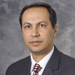
Mustafa Baskaya, MD
 Stroke is a major cause of morbidity and mortality in the U.S. population, particularly among aged individuals. Current demographic projections suggest that as the elderly population continues to expand, the need for developing improved treatment options for stroke patients will become even more pressing. Our lab is interested in the molecular events that occur during focal ischemia and in the use of stem cell approaches to improving neurological outcomes following stroke. Our main experimental model involves middle cerebral artery occlusion (MCAO) to generate focal ischemia in spontaneously hypertensive rats (SHR). Over the past several years, we have differentiated green fluorescent protein-expressing human embryonic stem cells (GFP-hESC) into mesenchymal stem cell-like cells (eMSCs). These eMSCs expressed markers (CD-29, CD-44, CD-73, CD-105, CD-166 and Nestin, but not CD-34, C45, CD-106, SSEA-4 or Oct-4) which are consistent with a mesenchymal stem cell state. We have transplanted eMSCs into the femoral veins of SHRs following MCAO. The expression of GFP allows us to follow the migration and further differentiation of the eMSCs in the ischemic brain. We observed migration of the injected eMSCs to the infarction region and their subsequent differentiation into neurons (which expressed β-tubulin III, MAP2, HuC and neurofilament) and vascular endothelial cells (which expressed vWF). The grafted cells do not appear to differentiate into astrocytes.
Nathaniel Brooks, MD
 The mission of the Surgical Interface Design Lab is to create and study tools that improve the ability of surgeons to perform progressively more complex procedures through more minimally invasive approaches. The lab does this by combining navigation, visualization and robotics technologies and refining the design to provide an intuitive, natural interface for the surgeon and the patient.
Robert Dempsey, MD
 Dr. Dempsey currently has multiple research projects with focus on cerebral ischemia and brain injury. As a Principal Investigator of grants from the National Institutes of Health, The Veterans Administration and the American Heart Association, Dr. Dempsey leads a group of neurosurgeons and scientists dedicated to neurosurgical patient care.
- The biochemistry of ischemic brain edema and traumatic brain injury.
- Lipid changes and other factors in the formation of carotid artery atherosclerosis.
- The stimulation and modification of adult neuro progenitor stem cells in brain after focal cerebral ischemia.
- Acute management of subarachnoid hemorrhage.
- Applied research in stroke, brain perfusion, subarachnoid hemorrhage and trauma.
- The molecular Biology of Embolic Carotid diseases and its relationship to delayed cognitive impairment.
Several clinical studies are ongoing including studies to look at the pathophysiology of a recurrent stroke, the possibility of supplementing blood flow to the brain after carotid occlusion, the impact of sleep disordered breathing on cerebral vascular regulation, and the modification of interoperative cerebral ischemia with hypothermia.
Amgad Hanna, MD
 Our lab is working on novel interventions to aid in functional and sensory recovery after spinal cord injury. Axons fail to regenerate appreciably after spinal cord injury resulting in patients losing both sensory and motor function below the level of injury. One of the primary reasons axons fail to regenerate is due to a glial scar that forms after an injury to the central nervous system. Our research uses peripheral nerve grafts to serve as scaffolds for axonal growth to bridge the glial scar. Peripheral nerve grafts provide several advantages when used as scaffolds; they contain Schwann cells that can myelinate regenerating axons as well as several growth factors that have been shown to promote axonal growth, and cytokines that modulate inflammation. To help enhance this axonal regeneration, we are also developing a sustained drug delivery platform to deliver therapeutic proteins at the graft-spinal cord interface.
Dan Hellenbrand, MS
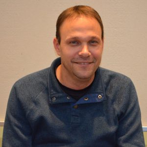 Dan works as an Assistant Researcher in Dr. Amgad Hanna’s Spinal Cord and Nerve Lab. He received his Bachelor of Science in Mechanical engineering Technology from Miami University in 2008 and a Master of Science in Biomedical Engineering from the University of Wisconsin in 2010. Dan’s primary research interests involve treatments to improve functional recovery after spinal cord injury. Dan, himself, sustained a C5-C6 spinal cord injury in 2003 and understand the hardships that result from the loss of motor and sensory function below the level of injury. In the lab, they are working on developing novel drug delivery methods to treat different aspects of spinal cord injury. The drug delivery platform is a mineral coating on surgical sutures or microparticles that binds specific therapeutic molecules of interest and releases them in a sustained manner. They are currently investigating the capabilities of interleukin-10, an anti-inflammatory cytokine, to reduce inflammation after spinal cord injury and the use of neurotrophin-3, a growth factor, to promote axon growth after spinal cord injury. For more information on our work and to view recent publications, visit Dr. Amgad Hanna’s page.
Bermans Iskandar, MD
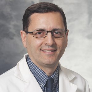 The role of folic acid supplementation has proven to be extremely effective in preventing the occurrence of neural tube defects and other congenital abnormalities in humans. Because this suggests that folic acid can modulate or enhance key mechanisms for growth and differentiation in the CNS, our laboratory has been instrumental in hypothesizing and proving a significant role for folic acid in regeneration and repair of the adult CNS after injury. As part of an effort to study the mechanism of such a pro-regenerative response, we have shown that folate-mediated CNS regeneration depends on injury-related induction of folate receptor FOLR1 expression, intact intracellular folate activation, and a functional methylation cycle. The effect of folate on the regeneration of afferent spinal neurons is biphasic and dose-dependent, and correlates closely over its dose range with global and gene-specific DNA methylation, and with expression of both the folate receptor FOLR1 as well as the de novoDNA methyltransferases. As such, we hypothesize an epigenetic mechanism in CNS repair, and through our current laboratory efforts, we seek to understand the underpinnings of this mechanism and its implications. This work provides possible avenues for new pharmacologic approaches to treating brain and spinal cord injuries.
Wendell Lake, MD
 Research support awarded and continued: Quantifying Changes in Neural Network Dynamics Elicited by Deep Brain Stimulation for the Treatment of Movement Disorders. MR Lease Account PRJ79UT Supported by the Departments of Radiology and Medical Physics, University of Wisconsin Awarded
Kip Ludwig, PhD
 Dr. Ludwig leads the Bioelectronic Medicines Laboratory at the University of Wisconsin, with the goal of developing next-generation neuromodulation therapies that use minimally invasive strategies to highjack the nervous system to treat circuit dysfunction and deliver biomolecules to target areas with unprecedented precision. Prior to Wisconsin Dr. Ludwig served as the Program Director for Neural Engineering at the National Institutes of Health. He co-led the Translational Devices Program at NINDS, led the NIH BRAIN Initiative programs to catalyze implantable academic and clinical devices to stimulate and/or record from the central nervous system, and led a trans-NIH planning team in developing the ~250 million dollar S.P.A.R.C. Program to stimulate advances in neuromodulation therapies for organ systems. Dr. Ludwig also worked in Industry as a research scientist, where his team conceived, developed and demonstrated the chronic efficacy of a next-generation neural stimulation electrode for reducing blood pressure in both pre-clinical studies and clinical trials. Through his industry work he oversaw Good Laboratory Practice (GLP) and non-GLP studies enabling clinical trials in Europe and the United States, as well as participated in the protocol development and execution of those trials, leading to approval for sale in seven countries and a U.S. Pivotal trial.
Suresh Mehta, PhD
 Suresh Mehta, PhD Profile
Gurwattan Miranpuri, PhD
Gurwattan Miranpuri, PhD, is a Senior Scientist working with Azam Ahmed, MD. Their work focuses on improving our understanding of stroke, both ischemic and hemorrhagic. In addition, Dr. Miranpuri is working on a research study, “Defining the epigenetic signatures of glioblastoma tumors and margins” in collaboration with Dr. Reid Alisch, PhD, Assistant Professor, Neurosurgery. This work is important in that it attempts to understand individual heterogeneity in the behavior of these aggressive tumors and may potentially lead to the development of important biomarkers. Together, and through this study, the research team has an extraordinary opportunity to characterize comprehensively, for the first time, glioblastoma tumors, as well as primary margins, to define molecular signatures of glioblastoma aggressiveness and margins. This study between basic scientists (Drs. Miranpuri and Alisch), neurosurgeons (Dr. Azam Ahmed) exemplifies collaboration within the department.
Eric Shusta, PhD
 The Shusta laboratory is focused on understanding the role of the brain vasculature, also known as the blood-brain barrier, in health and disease. To this end, they are designing new human induced pluripotent stem cell models of the diseased human blood-brain barrier to evaluate both disease processes and potential therapeutic treatments. They are also heavily invested in deploying state-of-the-art protein engineering tools to identify and optimize drug delivery strategies capable of targeted drug delivery to the central nervous system. These approaches rely on repurposing antibodies to act as targeting agents for the delivery of small molecules, biologics and nanoparticulate medicines. Taken together, these research thrusts promise to contribute to the diagnosis and treatment of debilitating neurological diseases.
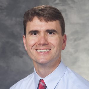
Aaron Suminski, PhD
 A fundamental problem in neural engineering and neuroscience is to understand how the activation of large populations of neurons gives rise to behavior at the level of the whole organism. For example, we often take the ability to move for granted because the complexities of interacting with our environment are masked by the reliability of motor behavior. This behavioral consistency is remarkable given the many factors that must be taken into consideration when moving the limb and manipulating objects, like the fidelity of sensory feedback, the goals of the task and the state of the limb. Our group aims to understand how neural activity at the ensemble and system levels produces the seemingly effortless dexterous control of the upper-limb despite the contextual complexity it involves using multiple methodologies including computational modeling, electrophysiology and functional brain imaging. Ultimately, we aim to translate our findings into the development of optimally effective therapies and technologies to improve the quality of life of patients with motor system dysfunction.
Raghu Vemuganti, PhD
 Noncoding RNAs and Post-Stroke Pathophysiology We are trying to understand the role of non-coding RNAs in brain damage following stroke and traumatic brain injury. Current projects are testing (a) if piRNAs altered after stroke leads to transposon dysfunction that is crucial for cell death, and (b) whether lncRNAs act as scaffolds for controlling gene expression after brain injury. MicroRNAs and Secondary Brain damage We are currently evaluating the role of several microRNAs in post-stroke pathophysiology. Particular projects include (a) evaluating how microRNAs and transcription factors control each other mutually after stroke, (b) deciphering the mechanism of microRNA-mediated gene induction in brain, (c) understanding the role of microRNA miR-29c in controlling DNA methylation and thus cellular homeostasis under ischemic conditions, and (d) evaluating if microRNAs control endoplasmic reticulum (ER) stress after stroke. Endoplasmic Reticulum Stress and Oxidative Stress in Neuronal Death We are currently testing if ER stress and oxidative stress are coincidental, potentiate each other bi-directionally and synergistically exacerbate the secondary brain damage after traumatic brain injury. Using a rodent model of controlled cortical impact injury, we are trying to answer the following questions. (1) What is the role of PERK-mediated ER stress pathway after TBI? (2) In the post-injury brain, are ER stress and oxidative stress connected? In particular, if ER stress mediated by PERK and oxidative stress modulated by NADPH oxidase NOX2 influence each other? (3) What is the effect of knocking-out/inhibiting individual rate-limiting proteins of PERK pathway eif2α, ATF4 and CHOP on oxidative stress and neuronal damage after TBI? Conversely, what is the effect of knocking-out/inhibiting NOX2 on ER stress and neuronal damage after TBI? The long-term goal is to understand the mutual interplay of ER stress and oxidative stress in post-trauma brain damage.
Uma Wesley, PhD
 The major goal of our research is to identify the therapeutic targets, and to elucidate the molecular mechanisms of post-stroke brain repair, tumor and stem cell survival, migration, and angiogenesis. We are particularly interested in the role of cell surface proteases and cytokines in regulating these physiological events that drive the post-stroke brain repair, and development and progression of cancers including glioblastoma/brain tumor. The soluble growth factors, cytokines, and extracellular matrix components (ECM) in the microenvironment contribute significantly to the stem cell dynamics, development of neuronal tumors, and brain injury repair following stroke. As a part of collaborative efforts with Dr. Dempsey our studies are expected to bridge tumor cell studies to stem cell regulated repair of cerebro-vascular diseases, particularly ischemic stroke. Projects include (1) Understanding the biochemical and molecular mechanisms of protease and chemokine regulated cellular functions; (2) Regulation of cytokine/chemokine signaling pathways during tumor development and brain injury repair; (3) Identifying and targeting molecules involved in progression of atherosclerotic plaques associated with ischemic stroke. Our research utilizes interdisciplinary approaches that include in vitro cell culture, and in vivo rodent models; Biochemical, cellular and molecular biology approaches, immune-fluorescence microscopy, protein analysis/proteomics/protein array screening. Funding: Dr. Wesley has successfully obtained research/grant funding as a principal investigator from National Institute of Health (NIH), American Heart Association (AHA), and Wisconsin Women’s Health Foundation (WWHF), and UW-Carbone Cancer Center.
Justin Williams, PhD
 The Neural Interface Technology Research and Optimization (NITRO) Laboratory is part of the Department of Biomedical Engineering at the University of Wisconsin-Madison. The goals of our lab are to 1) develop new devices for recording from and stimulating neural tissue, 2) design these devices to be both durable for long-term implantation and safe for use in humans and animals, and 3) use these technologies in a variety of situations, from use in a basic physiology lab recording from single neurons, to clinical settings where people with motor disabilities might benefit from a brain-computer interface or other neural prosthetic communication device.